Photos of the microscopic show us that indeed the universe is a wondrous place. Artistic complexity and awe-inspiring design underlie the smallest detail beyond our eye’s ability to see. Nikon’s annual Small World Photomicrography Competition is judged for both artistry and scientific technique and if you like the examples below there are thousands more to explore at Nikon’s fabulous website.
My favorite below is the fruit fly sperm. What about you?
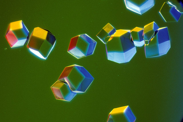
Crystals of influenza virus isolated from terns by Julie Macklin and Graeme Laver, Canberra, Australia
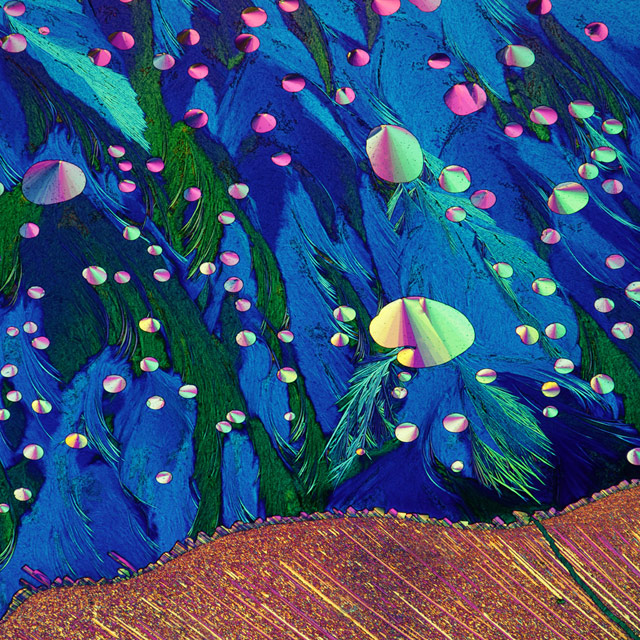
Crystals of sulphur and other acids by Dr. John, the Department of Atmospheric and Oceanic Sciences
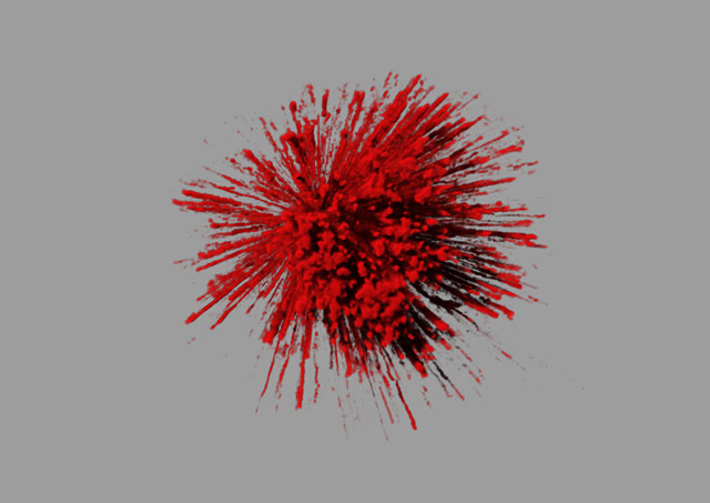
The explosive dynamics of sugar transport in fat cells, Sydney, Australia
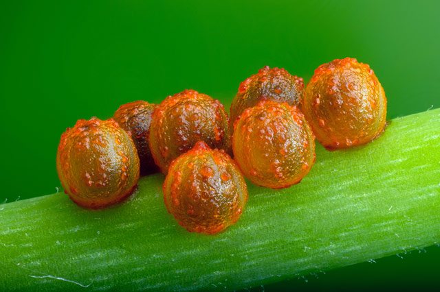
Butterfly eggs on a stem, by David Millard, Austin, Texas
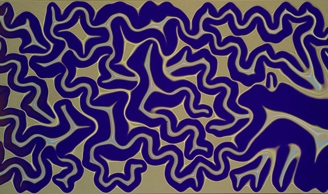
Silicon dioxide on polydimethylglutarimide-based resist, Canada by Dr. Pedro Barios Perez,
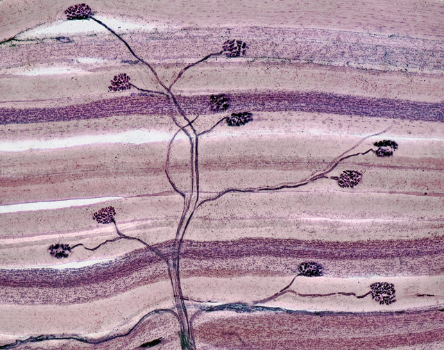
Nerve and muscle thin section, by David Ward, Oakdale, CA
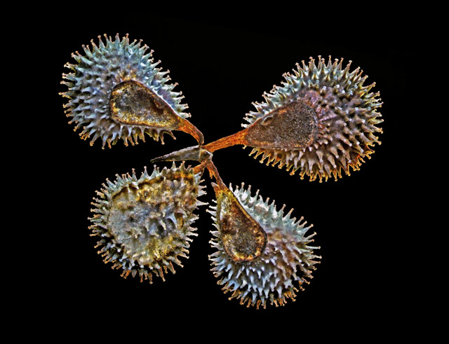
Gypsy flower seeds by Keszthely, Zala, Dr. Csaba Pinter, Hungary
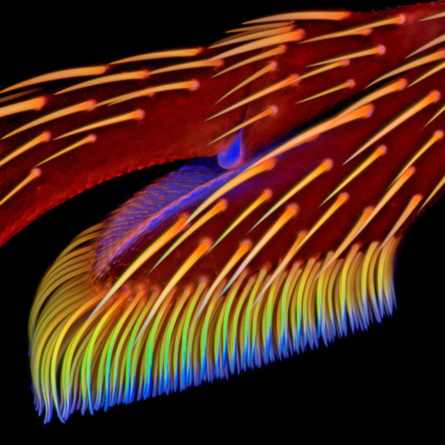
Adhesive pad on a foreleg of a ladybird beetle, by Jan_Michels.
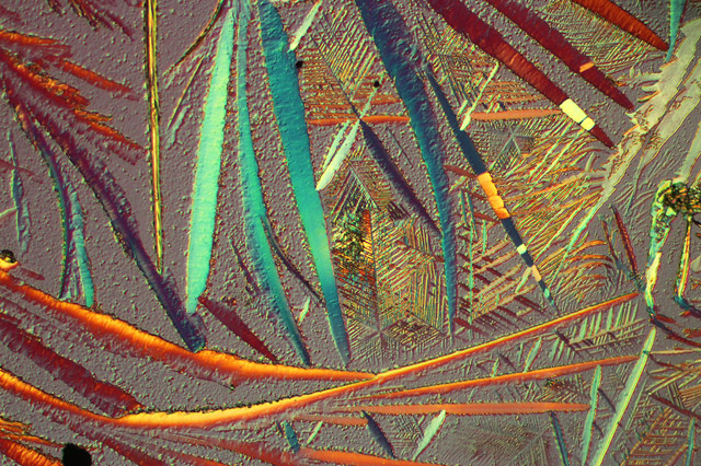
Battery leakage crystal, National Astronomical Observatories, Chinese Academy of Sciences, Zhang Chao
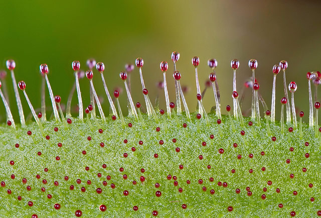
Glands of a leaf by University of Puerto Rico (UPR), Mayaguez Campus, Jose Rivera

Vitamin C crystal by Raul Gonzalez Estudio
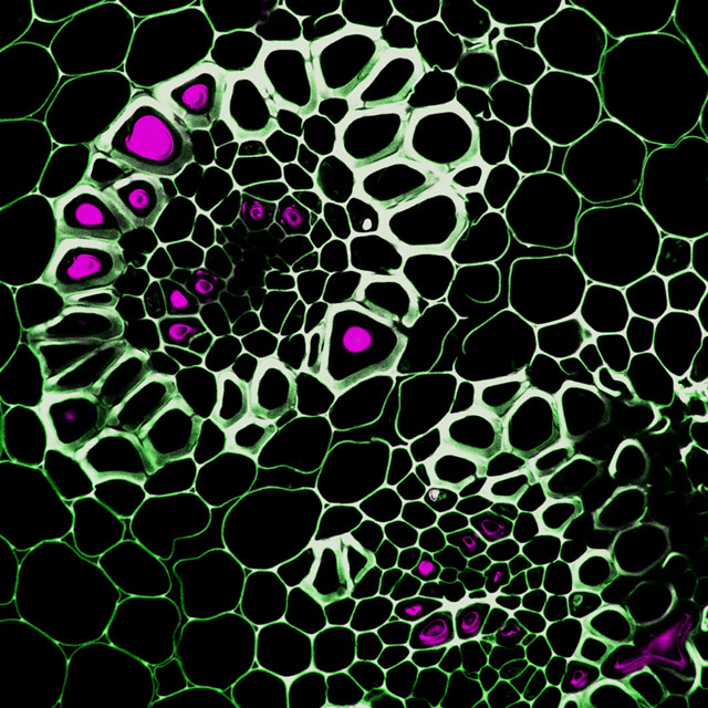
Lily of the Valley, rhizone section by Dewel Microscopy Facility, Dr. Guichuan Hou,
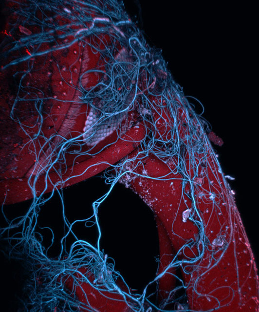
Insects wrapped in spider web, by Mark Sanders, minnesota
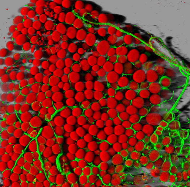
Mouse fat tissue, National Heart, Lung and Blood Institute, Light Microscopy Core Facility, Dr. Daniela Malide
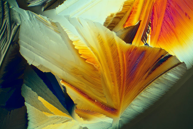
Citric and tarteric acid by Herald Anderson, Steinberg, Buskerud, Norway, Herald Anderson
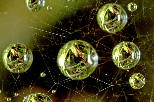
Dew on a spider web by Massimo Brizze, www.massimobrizzi.it
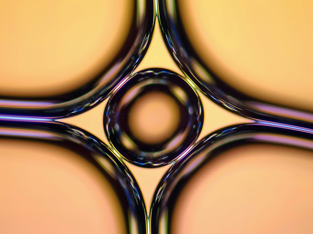
Soap bubble, by Haris Antonopoulo, Athens, Greece
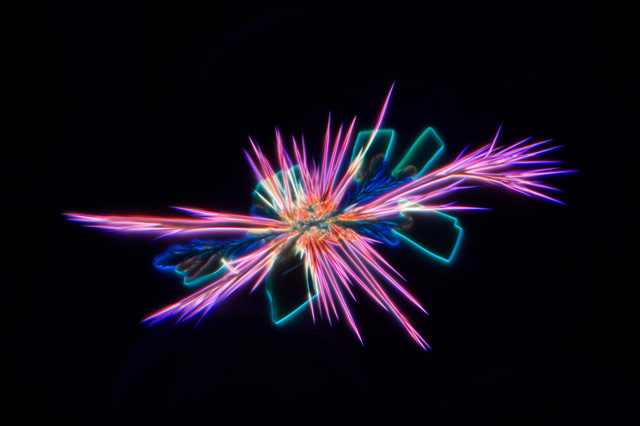
Purple food dye, by Waldo Nell, Surrey, British Columbia, Canada
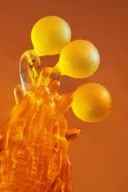
Pollen-crocus,by Frederic Labaune, Auxonne, France
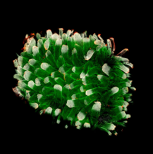
Small intestine of a mouse by Dr. Bryan Millis, National Institute on Deafness and Other Communication Disorders, Laboratory of Cell Structure and Dynamics
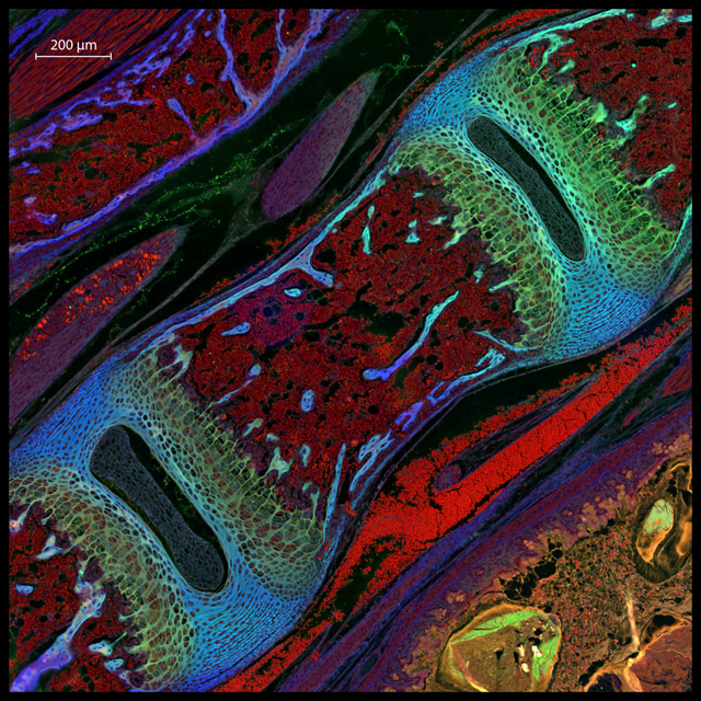
Mouse vertebrae section by Michael Nelson et al, Alabama, US
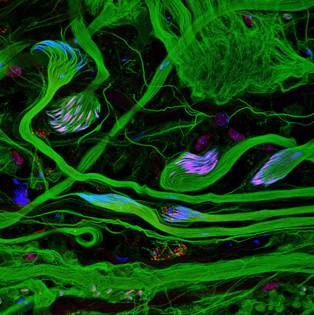
Fruit fly sperm by Dr. Janet Rollins, College of Mount Saint Vincent, New York
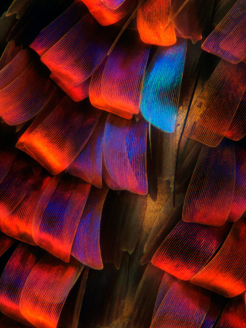
Madagascan Sunset moth by Maidstone, U.K., Laurie Knight
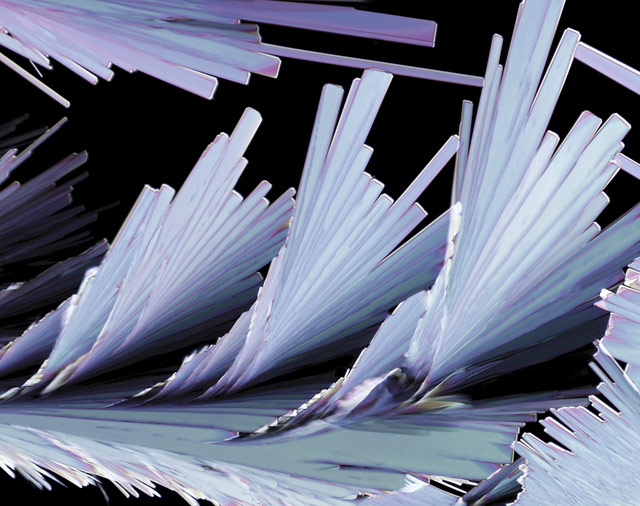
Sulfosalicylic Acid Crystals,by Thomas Balla
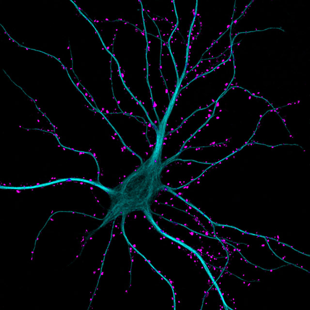
Hippocampal neuron receiving excitatory contacts, by Dr. Kirian Boyle, glascow

paramecium
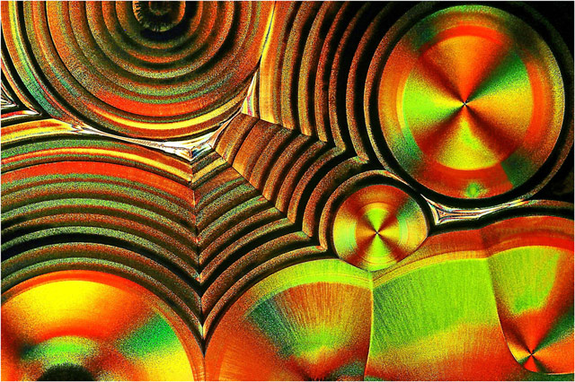
Rings and Circles, Vitamin-C Microcristals, by Rudolf Bauer
Here’s a chance to see more, first at this post, then the Nikon site again.


Just awe-inspiring! Everything was designed beautifully….
Fabulous images!
breathtaking…..how can anyone ever be bored? Thank you for these wonderful pictures Dusky.
Cecilia
these images transport one to a different world; microscopic OR NOT…They really open-ones-eyes TOO….
Amazing…ck.-out the small intestine of a mouse, as well as some of the other vibrant & colorful images…It’s also gonna take me awhile to take my vitamin C tablets….
Wow! The newspapers are reporting that vitamins are worthless, via the AMA and traditional medical journals. How can that Vitamin C crystal NOT do something helpful to the body?
What nature offers, amazing!
simply gorgeous!
What a wonderful world, indeed.
all of these pic’s are just soooo awesome i had to look at them over and over again…
thank you so much.
patti
These are just fantastic! I can’t pick a favorite, they are all so wonderful. I love looking at photographs of microscopic things. They are beautiful in an almost other-wordly way.
All the photographs prove that the God is the greatest artist. look at the colors and composition, no one can beat HIM even at the microscopic level.
My choice is the sulfur crystals as it reminds me of Peacock feature
Just fantastic , more and more
I love this. Seeing parts of things you sometimes hear about but never see. I want to see all you have to show.. But then how do I find the new stuff, can I keep the older stuff so I can go back and see it again? I love looking and watching.
Just wonderful ,looking forward to my other Serch for a Wonders God bless from Margaret Clancy
Check this out